
CBCT & 3D Dentistry
Advanced 3D Imaging for Oral Surgery Planning
What Is CBCT Imaging?
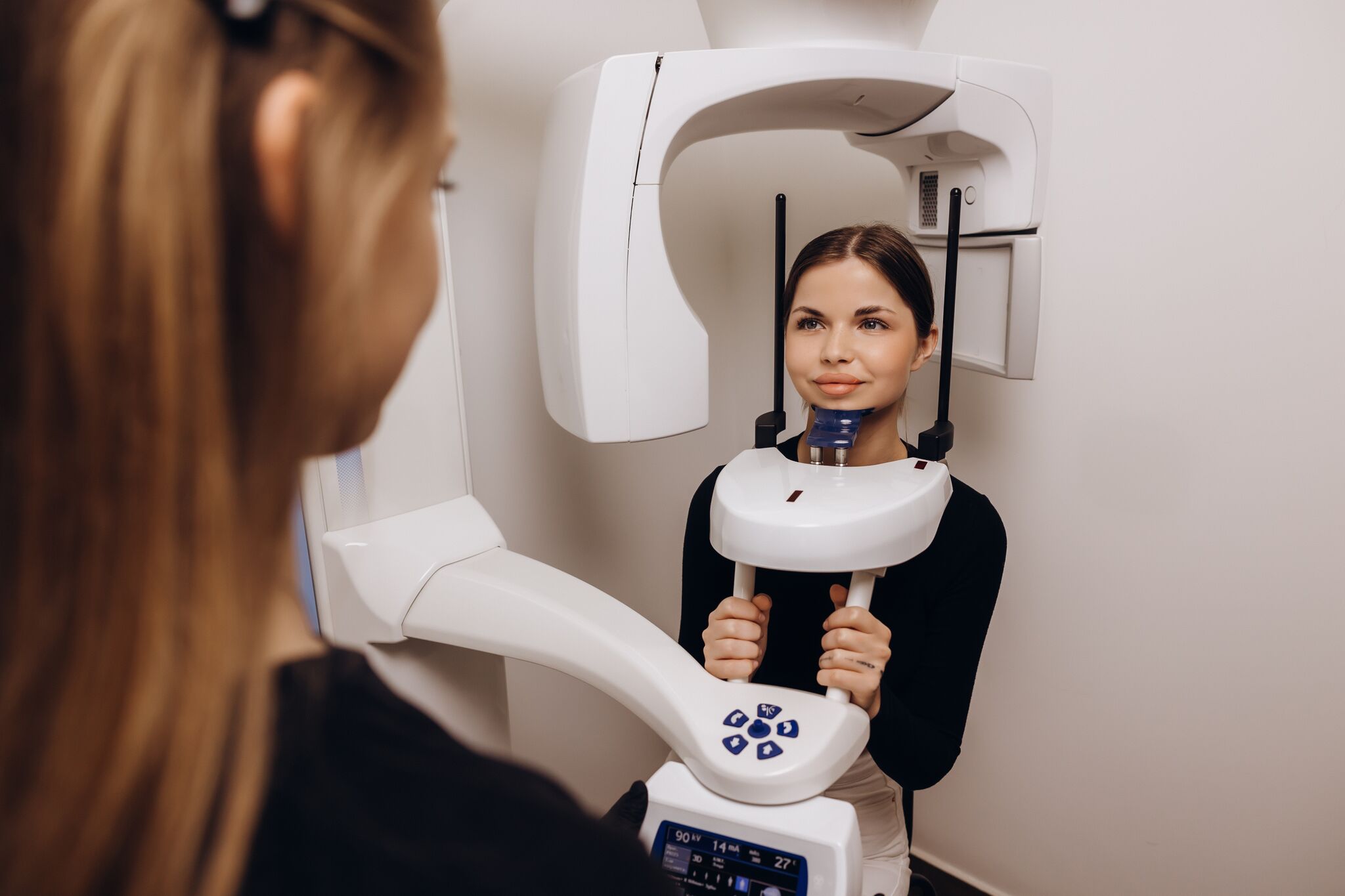
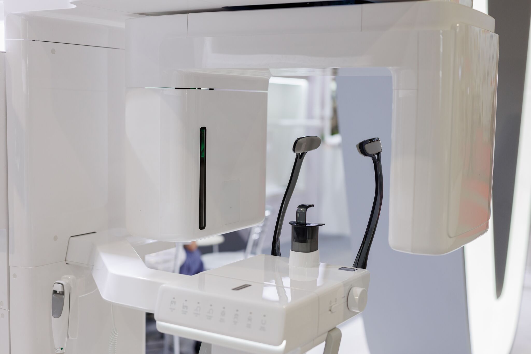
Cone beam computed tomography, commonly known as CBCT, is an advanced imaging technology that creates detailed three-dimensional images of the teeth, jaws, and surrounding structures.
Unlike traditional dental X-rays, which produce flat two-dimensional pictures, a single CBCT scan shows bone, tooth position, nerves, and sinuses from multiple angles.
At Desert Oral Surgery, 3D imaging may be used to support diagnosis and treatment planning for dental implants, wisdom teeth removal, impacted canines, jaw surgery, and other oral and maxillofacial procedures.
Who It Helps
When 3D Imaging May Be Recommended
3D imaging may be recommended when your oral surgeon needs a more detailed view before treatment, including:
- Dental implant planning
- Bone grafting evaluation
- Wisdom teeth evaluation
- Locating impacted canines
- Jaw surgery planning
- Facial trauma assessment
- Oral pathology evaluation
Your oral surgeon will determine whether a CBCT scan is needed based on your examination and the procedure being planned.
What to Expect
How 3D Imaging Works
-
Imaging Evaluation
A 3D scan may be taken during your visit when additional detail is needed to understand your anatomy and plan treatment.
-
Digital Review
The scan produces a three-dimensional image that your surgeon can rotate and examine digitally to evaluate bone structure, tooth position, nerves, sinuses, and other important areas.
-
Treatment Planning
Your surgeon uses the image to plan the procedure in detail, including implant position, surgical access, grafting needs, and protection of nearby structures.
-
Personalized Surgical Care
The completed plan guides your procedure and helps your surgeon explain the recommended approach to you before treatment begins.
Because the scan shows your anatomy in three dimensions, it helps your surgeon anticipate the details of your case before the procedure.
Imaging That Guides the Procedure
Your surgeon reviews the completed scan with you and explains what it shows about your anatomy before any treatment is planned.
Seeing the bone, tooth position and nearby structures together makes it easier to discuss the recommended approach and what to expect.
Benefits of 3D Dentistry
Why 3D Imaging Matters
3D imaging gives the surgical team a more complete view of your anatomy, which supports careful planning and informed clinical decisions. Potential benefits include:
- More detailed diagnosis
- Clearer visualization of bone, teeth, and soft tissue relationships
- Improved dental implant planning
- More precise evaluation of impacted wisdom teeth and canines
- Better understanding of nerve and sinus location
- Support for conservative surgical planning
- Clearer patient education before treatment
Your oral surgeon will review your images with you and explain how they inform your treatment plan.
Location
Serving Palm Desert & Palm Springs
Desert Oral Surgery uses 3D imaging to support oral surgery evaluations and treatment planning for patients in Palm Desert and Palm Springs.
-

Palm Desert Office
- Address: 72-780 El Paseo, Suite E-1, Palm Desert, CA 92260
- Phone: (760) 346-7431
- Hours: Mon to Fri: 8:00 AM to 5:00 PM · Sat and Sun: Closed
-

Palm Springs Office
- Address: 1900 E Tahquitz Canyon Way, Suite A-1, Palm Springs, CA 92262
- Phone: (760) 327-1509
- Hours: Mon to Fri: 8:00 AM to 5:00 PM · Sat and Sun: Closed
Related Services
Related Oral Surgery Services
Get Started
Learn More About 3D Imaging for Your Treatment
If you need dental implants, wisdom teeth removal, bone grafting, jaw surgery, or another oral surgery procedure, 3D imaging may help your surgeon develop a clearer and more precise treatment plan. Desert Oral Surgery can evaluate your needs and explain whether CBCT imaging is recommended for your care.